
Researchers at the University of Basel dissected twelve cadaver heads and cross-checked the tissue against CT and MRI scans, looking closely at the masseter muscle that closes the jaw during chewing.
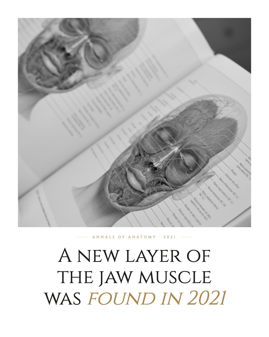
Underneath the two known layers, the team found a third, a distinct band of muscle fibers running from the zygomatic arch to the coronoid process of the jawbone, which they named the masseter pars coronoidea. Its fiber direction differed from the other layers, suggesting it pulls the jaw in a slightly different way during biting and clenching.
Because the masseter is one of the most frequently imaged and operated-on muscles in the face, from jaw surgery to Botox injections for clenching, a missing layer may matter for how those procedures are planned. Only a few historical texts had hinted at a third layer, and they disagreed on where it lay.
Read the original study
Mezey et al., Annals of Anatomy, 2021 · doi.org/10.1016/j.aanat.2021.151879
A chewing muscle hid an extra layer.
Source
- Mezey, S. E., Müller-Gerbl, M., Toranelli, M., & Türp, J. C. (2022). The human masseter muscle revisited: First description of its coronoid part. Annals of Anatomy = Anatomischer Anzeiger, 240, 151879. https://doi.org/10.1016/j.aanat.2021.151879