
Researchers at Heinrich Heine University in Germany noticed that some of their brain organoids were spontaneously forming small, symmetric structures on their surface, and used imaging and gene analysis to find out what the structures actually were.
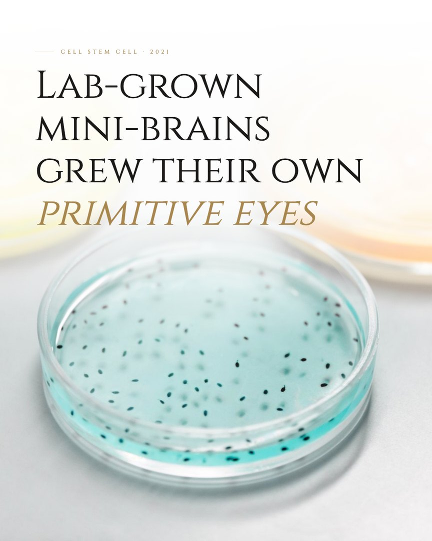
By about 60 days of growth, the organoids had formed paired, functionally integrated optic vesicles, the structure the eye's retina develops from in an embryo, complete with retina-like tissue, lens-like cells, and neurons that responded to changes in light intensity.
The organoids were growing a piece of early eye anatomy without being instructed to, simply because the same developmental program that builds the eye is wired into the same stem cells that build the brain. The structures were primitive and not functional vision, and the authors proposed using them to study how eye and brain tissue develop together.
Read the original study
Gabriel et al., Cell Stem Cell, 2021 · doi.org/10.1016/j.stem.2021.07.010
A brain in a dish, growing its own eyes.
Source
- Gabriel, E., Albanna, W., Pasquini, G., Ramani, A., Josipovic, N., Mariappan, A., Schinzel, F., Karch, C. M., Bao, G., Gottardo, M., Suren, A. A., Hescheler, J., Nagel-Wolfrum, K., Persico, V., Rizzoli, S. O., Altmüller, J., Riparbelli, M. G., Callaini, G., Goureau, O., . . . Gopalakrishnan, J. (2021). Human brain organoids assemble functionally integrated bilateral optic vesicles. Cell Stem Cell, 28(10), 1740–1757.e8. https://doi.org/10.1016/j.stem.2021.07.010